

By Dr Oliver Liyou BVSc (Hons) MACVSc
Published in Australian Stock Horse Journal July-August 2004
It is not uncommon in practice to be asked to examine a horse for a lump on the head of a horse. The inability of the horse to inform us how and when the lump started can make diagnosing the cause of these a challenge at times.
As with most medical problems, it is only with a correct diagnosis that the correct treatment can be instigated. Sometimes other signs of disease elsewhere in the body, eg. lungs, may assist in reaching a diagnosis, and so a complete clinical examination is necessary.
A thorough history taking is also important eg when the lump was first noticed, is it painful, is it increasing or decreasing in size etc. You as the owner will be asked questions like — "is it affecting the horse's attitude, how is the horse eating etc".
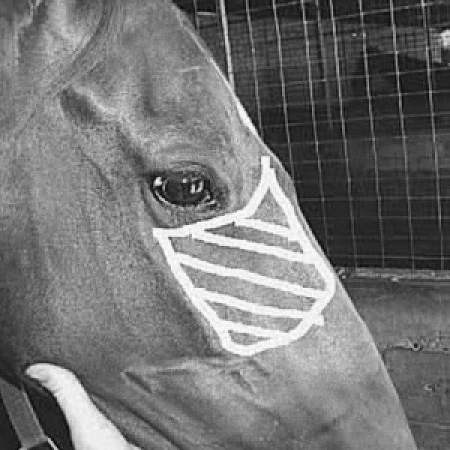
Close inspection of the head region is the next step in reaching a diagnosis of the lump's cause.
The external palpation will check things such as:
- symmetry of the head and jaw — is one side larger, flatter etc?
- condition and health of the skin near lesion and rest of head eg is there hair loss, signs of itching etc?
- is there heat and/or pain over the lump?
- is there any fluid discharge from the lump eg pus?
- is the lump hard or soft, round or long and irregular etc?
- is the lump in the skin and so freely moveable from the underlying bone etc?
- the position of the lump and what structures is it near eg is it at the site of the root of a tooth?
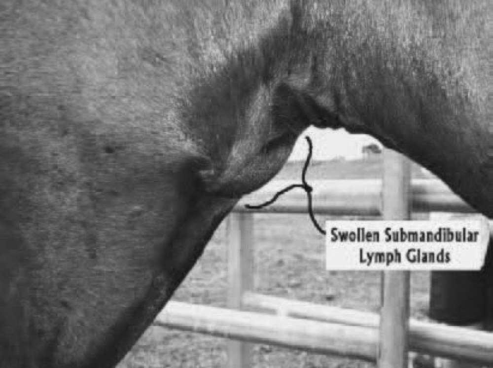
The lymph nodes (the glands under the throat), which are important in fighting infection, may become enlarged if infection is present in the head region. This infection could be bacterial, viral or fungal in nature.
The salivary glands too can become involved and so need to be inspected.
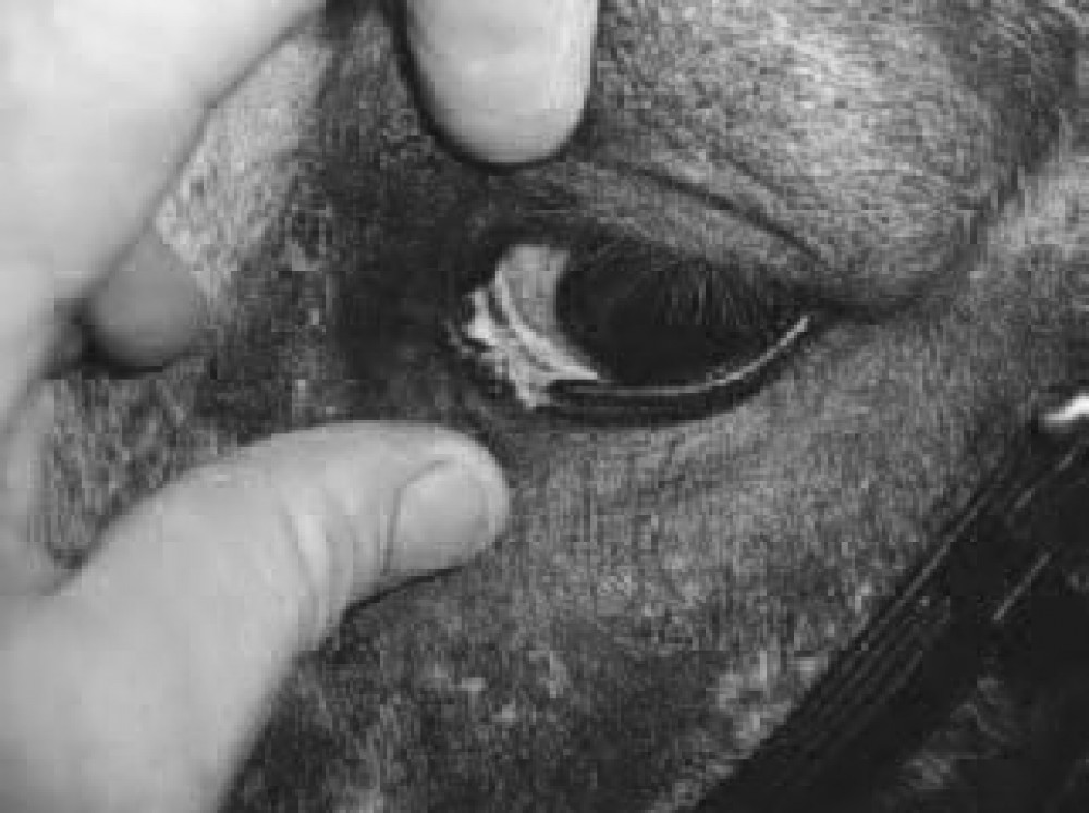
The eyes, ears and nostrils may show altered function eg discharges, squinting with pain, flaring with certain conditions eg infections, trauma, signs of cancer etc and so need to be closely examined.
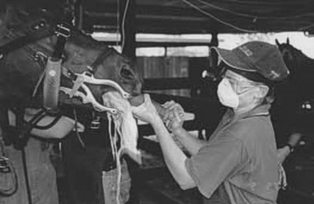
An oral examination is paramount in investigating lumps which could be involving the mouth. Examination can be enhanced by using a full mouth speculum, flushing out the mouth and the use of a light source.
Further investigation of head lumps is often required. This may involve radiographs (x-rays), endoscopy, ultrasound etc.
Some of the possible causes of head lumps will be listed below and then discussed briefly:
- teething bumps (eruption cysts) in 2-5 year old horses
- impacted teeth
- abscessed teeth
- quidding or balling of feed in the cheeks whilst eating
- cysts in nasal cavity, skin etc
- foreign body eg grass seed abscess
- soft tissue swellings eg bites, allergic reactions
- hamartoma and teratoma (developmental abnormalities)
- cancers of the head, nasal cavity, mouth and skin eg sarcoids, sun cancers, melanomas etc.
- ethmoidal haematomas (blood tumors in the rear part of the nasal cavities)
- "big head" (nutritional hyperparathyroidism)
- bone spurs
- suture periostitis
- blunt trauma
- sinusitis (inflammation in the sinus cavities)
- hypertrophic osteopathy from lung disease
- other conditions are seen but are too numerous to list fully here.
Some of the causes of lumps found on horses' heads are related to the teeth.
Teething bumps (eruption cysts) are the most common lumps seen in young horses. These are caused by the pressure effects of the permanent tooth (which is still under the gum line) trying to "push out" the deciduous teeth (caps or baby teeth). If these bumps are not painful or hot, then they are usually not a problem. However they are a reminder that young horses may need frequent dental check ups eg as often as every 3-6 months in the 2.5 — 4 year old period.
Impacted teeth, tooth root abscesses and quidding of food are due to periodontal disease etc.
Sinus cysts are fluid filled cavities which develop in the sinuses next to the nasal cavity. Thus they will cause a diffuse facial swelling on one side of the face — in an area below the eye and half way to the nostril. This area represents where the maxillary sinus is positioned in the skull.
The cause of sinus cysts is unknown. They usually present as a facial swelling on one side which progressively gets bigger. A clear discharge from the nostril on the swollen side is commonly reported. The swelling may get so big that it affects the ability of the horse to breathe properly during exercise.
Sinus cysts can occur in horses under 1 year of age or horses over 9-10 years of age. X-rays and possibly the use of an endoscope are necessary to confirm the diagnosis. Surgery is the required treatment for this condition.
Another cause of sinus swelling is sinusitis which is where the sinus becomes infected and a lot of pus is produced in the cavity. This pus may build up and thus cause major facial distortion, or continue to drain well from one nostril — resulting in little facial distortion. Tooth root abscesses which drain into the sinuses are reported to cause little in the way of facial distortion.
Cancers of the nasal cavity and sinuses can also cause distortion to the face.
Soft tissue swellings such as bites and allergies are common causes of lumps on horses' heads. Infections and abscesses from foreign bodies are also encountered frequently. Because horses lead with their head when grazing, snakes usually strike at the head or the legs of the horse. Some snake bites result in little facial swelling, but others can cause considerable swelling of the affected area, and sometimes massive swelling of the large masseter muscles on the cheeks.
Abscesses can occur from grass seeds penetrating the skin, and also from cuts inside the mouth from sharp teeth. The cuts and foreign body penetration lead to infection of the cheeks which can lead to abscess formation. As large amounts of pus form in these abscesses, skilled surgical draining is often necessary. Antibiotics, anti-inflammatory and tetanus coverage etc may also be necessary. Skill of the surgeon is crucial as there are many important blood vessels, nerves, salivary ducts etc which traverse parts of the cheek region, and these must be avoided in the surgical incisions.
Some soft tissue swellings of the head can continue to swell and become medical emergencies, as they can begin to cut off the airways.
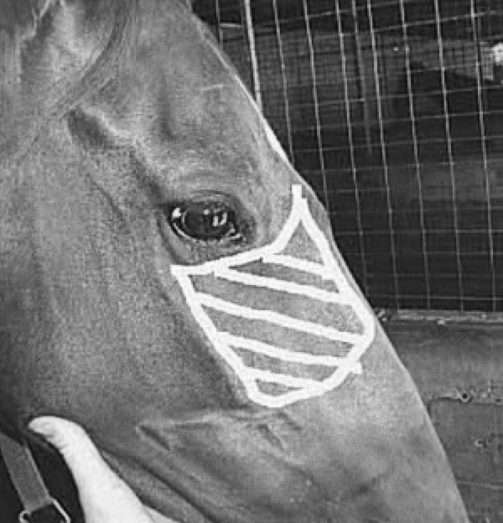
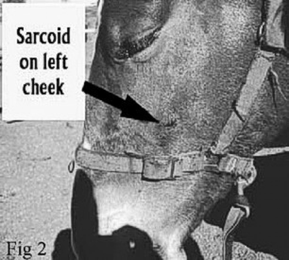
Cancers of the head's skin and subcutaneous tissues are commonly encountered. Sarcoid tumors, squamous cell carcinomas and melanomas of grey horses are seen often. These tumors can at times be malignant and may enlarge or spread to other sites in the body. Thus early diagnosis, and possibly treatment, by a veterinarian is essential for a successful outcome.
"Big head" or nutritional secondary hyperparathyroidism (NPH) is a condition whereby the horse's body is not receiving enough calcium. This calcium deficiency may be the result of the horse eating grasses which are high in oxalates. The oxalates bind to the calcium and render it not able to be absorbed by the horse's gut. Buffel grass, kikuyu and setaria are the most common grasses to cause big head.
As most grains are low in calcium and high in phosphorus, a high grain diet needs to be supplemented with calcium to be balanced, and thus avoid the risk of big head.
Big head may present as a shifting lameness (whereby the horse's lameness changes from one leg to another from time to time), and the horse may become reluctant to move. As the bones are weaker, they are at risk of fracturing and so the affected horse should not be ridden.
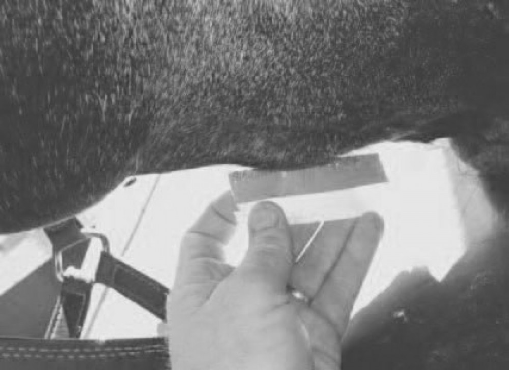
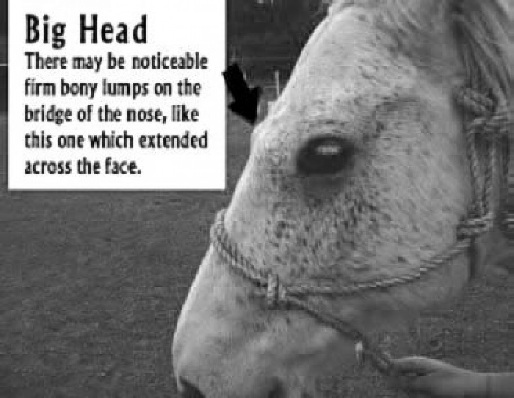 Above: an example of "big head"
Firm bony swellings of the bridge of the nose may be a sign of big head. It is important to note that there are many other causes of firm bony lumps on the bridge of the nose, so investigation by a veterinarian is important here.
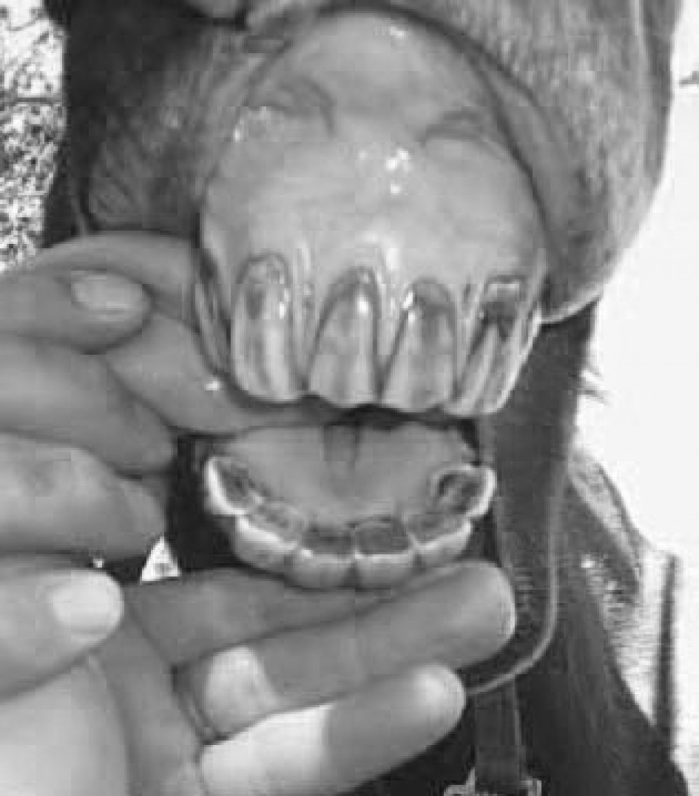
With big head, the jaw bones (mandible rami) may become noticeably thicker when felt from underneath the jaw. Sometimes the bones of the mandible (lower jaw) become so thick and distorted that the tongue hangs out, and the front incisor teeth cannot meet. Confirmation of the diagnosis may require a blood and urine test to measure calcium excretion from the body whilst on that particular diet.
Blunt Traumas such as kicks to the head, running into a tree limb etc may cause firm bony lumps which appear similar to big head, especially if they are not noticed early.
Any lump which is painful and has appeared suddenly may be suspected of being of traumatic origin. The bony swelling often reduces in size, but this can be a slow process.
Suture periostitis is a condition similar to "splints" in the legs. It is where there is a lot of bony inflammation along the suture lines of the nasofrontal region. The suture lines are the borders of the individual bones of the skull. If these become traumatised, twisted etc, then bony reaction and thickening occurs along them. Thus an appearance similar to big head and blunt trauma will result. Here, x-rays can help to confirm the diagnosis.
It becomes obvious that there are many varying causes of lumps and swellings on horses' heads. Thorough investigation is important to differentiate what the cause is, so a diagnosis and the best treatment, if necessary, can be implemented as soon as possible.